Probiotics for Mucus in Stool: What the Science Says About Restoring the Gut's Mucosal Barrier
Understanding the connection between gut bacteria, the intestinal mucus layer, and what it means when that balance breaks down
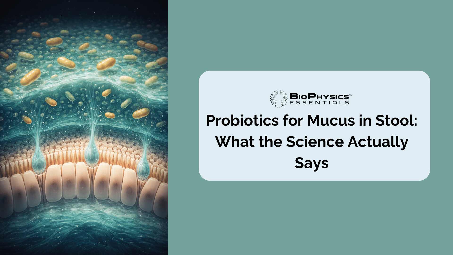
Seeing mucus in your stool is one of those symptoms that tends to get dismissed—or, on the other end, cause immediate alarm. The truth is somewhere in between. A small amount of mucus is a normal byproduct of healthy digestion; it's produced continuously by goblet cells lining the intestinal wall to lubricate the passage of stool and form a protective barrier between gut contents and the underlying tissue. But when mucus becomes visible, persistent, or unusually heavy, it's a signal that the mucosal layer is under stress.
What fewer people realize is that the gut microbiome plays a central role in regulating this mucus system. The same probiotic bacteria that support digestion, immunity, and inflammation control are also directly involved in the production, maintenance, and composition of the intestinal mucus layer. When the microbial community shifts—through dysbiosis, antibiotic use, infection, or chronic inflammation—the mucus layer can thin, become disrupted, or produce excess mucus in response to irritation.
This article examines the peer-reviewed evidence on how specific probiotic strains interact with the gut mucus barrier, which conditions most commonly drive increased mucus in stool, and what the research tells us about using probiotics to address the underlying imbalances. Because this isn't primarily about managing a symptom—it's about restoring the gut environment that keeps that symptom from appearing in the first place.
Key Takeaways
- Mucus in stool is a sign of mucosal stress. The intestinal mucus layer protects the gut lining. When visible mucus appears in stool, it often reflects an underlying disruption—inflammation, infection, dysbiosis, or barrier breakdown—rather than a standalone symptom.[1]
- Probiotic bacteria directly regulate mucin production. Strains like Lactobacillus plantarum and L. rhamnosus have been shown to upregulate MUC2 and MUC3 mucin gene expression in intestinal epithelial cells, strengthening the layer that keeps gut contents separated from immune tissue.[2]
- Lactobacillus reuteri significantly increases mucus layer thickness in preclinical models of colitis, while also reducing key inflammatory markers including IL-1β and IL-6.[3]
- Bifidobacterium longum restored the mucus barrier in animal studies of Western diet–induced dysbiosis, with supplementation reducing mucus degradation and re-establishing barrier function.[4]
- Multi-strain Bacillus probiotics reduced fecal mucus in children with persistent diarrhea significantly faster than standard care alone, with the probiotic group achieving mucus resolution in a median of 2.6 days vs. 5.0 days in controls.[5]
- Probiotic mucus adhesion predicts colonization effectiveness. L. rhamnosus GG demonstrated 34% adhesion to intestinal mucus—the highest of five tested strains—suggesting superior persistence and mucosal influence compared to lower-adhering competitors.[6]
What Mucus in Stool Actually Means
The intestinal mucus layer is not a passive feature of gut anatomy—it is an active, continuously renewed biological barrier that plays a foundational role in gut health. Produced by specialized goblet cells throughout the colon, the gel-forming protein MUC2 is the primary structural component of this layer. It forms a bilayer system in the colon: a sterile inner layer tightly adhered to the epithelium, and a looser outer layer that the microbiome can interact with.[1]
Under normal conditions, mucus is continuously secreted and shed into the lumen, meaning small quantities routinely mix with stool. This is entirely normal and typically not visible. What changes this picture is the word visible—when mucus appears as white, yellow, or clear strands in the toilet bowl or on stool, the volume has increased meaningfully, or the mucus composition has changed in response to an irritant.
What Triggers Excess Mucus?
When the intestinal mucosa is irritated or inflamed, goblet cells respond by increasing secretion. This is a protective reflex—more mucus means more barrier protection. But it's also a diagnostic signal. Research consistently links visible mucus in stool to the following categories of underlying cause:[7]
- Irritable Bowel Syndrome (IBS): Mucus in stool is a recognized symptom across IBS subtypes, particularly IBS-D and IBS-M, affecting a meaningful proportion of patients. The gut-brain axis, motility changes, and mild immune activation can all drive mucus secretion without visible inflammation on colonoscopy.
- Inflammatory Bowel Disease (IBD): Both ulcerative colitis and Crohn's disease are associated with mucus—sometimes blood-tinged—due to active mucosal inflammation and ulceration. Mucus in these conditions reflects direct damage to the epithelium.[8]
- Gut Dysbiosis: Disruption of the microbial community can thin the mucus layer and alter goblet cell function, creating a cycle where barrier breakdown leads to more inflammation and further mucus secretion.
- Post-infectious gut changes: Bacterial gastroenteritis, Clostridioides difficile infection, and other enteric infections can trigger acute mucus production and, in some cases, persistent post-infectious IBS with ongoing mucosal symptoms.
- Antibiotic-associated dysbiosis: Broad-spectrum antibiotics reduce microbial diversity and can deplete the species that sustain mucus layer integrity, leaving the gut lining temporarily vulnerable.
Important: When Mucus Requires Medical Evaluation
Mucus alone—particularly if clear or white and without blood—is often a benign finding in the context of IBS or mild dietary disruption. However, mucus accompanied by blood, significant abdominal pain, fever, unexplained weight loss, or a change in bowel habits that persists beyond a few weeks warrants evaluation by a gastroenterologist. These combinations can be associated with IBD, infection, or, less commonly, colorectal cancer. The information in this article is not a substitute for medical diagnosis.
The Gut Microbiome and Your Mucus Layer

The relationship between gut bacteria and the mucus layer runs in both directions. The mucus layer provides a habitat and nutrient source for commensals, particularly in the outer layer. In return, healthy commensal bacteria—especially Lactobacillus and Bifidobacterium species—actively support mucin gene expression, goblet cell function, and the structural integrity of the barrier itself.[9]
The Science of Mucin Production
The intestinal mucus layer is primarily composed of MUC2, a large, heavily glycosylated protein assembled by goblet cells and secreted into the lumen. Mucus is not static—it's continuously produced and shed, creating a flowing barrier that moves outward from the epithelium. When the microbiome is in balance, commensal bacteria stimulate appropriate mucin gene expression. When dysbiosis occurs, mucin-degrading species like certain Bacteroides and Akkermansia strains (which in excess can erode the mucus layer faster than goblet cells can replenish it) may expand, thinning the protective barrier and triggering the inflammation that drives visible mucus in stool.[10]
This is why addressing mucus in stool through microbiome support—rather than simply trying to suppress the symptom—reflects the underlying biology more accurately. The bacteria that bind to mucosal surfaces are particularly important here: strains with demonstrated mucus adhesion properties form a physical colonization layer that competes with pathogens, supports goblet cell signaling, and helps regulate the entire barrier ecosystem.
When antibiotic use, poor diet, or infection disrupts this ecosystem, the consequences can include both an acutely thinned mucus layer (increasing permeability and immune activation) and, paradoxically, a reactive over-secretion of mucus as the gut attempts to compensate. Both scenarios can manifest as visible mucus in stool.
Probiotic Strains With Evidence for Mucosal Support
Not all probiotic strains interact with the mucus layer equally. The following strains have peer-reviewed evidence supporting their role in MUC2 upregulation, mucus layer thickness, mucus adhesion, or mucosal inflammation reduction. All strains discussed below are present in MicroBiome Restore.
Lactobacillus plantarum: MUC2 Upregulation and Barrier Fortification
Lactobacillus plantarum has one of the strongest track records in mucin research among Lactobacillus species. A randomized, double-blind, placebo-controlled trial published in Scientific Reports found that L. plantarum TIFN101 upregulated the MUC2 gene in small intestinal mucosal tissue. Mucin 2 is produced by goblet cells and forms the primary scaffolding of the protective mucus gel layer—its increased expression directly translates to a stronger barrier.[11]
Additional research demonstrated that L. plantarum PS128 significantly increased the amount of colonic mucin in mice, including in a constipation model where mucus had been experimentally reduced. Immunohistochemical analysis confirmed higher MUC2 protein levels in both standard and loperamide-treated mice receiving this strain.[12] In vitro research further showed that L. plantarum 299v and L. rhamnosus GG both upregulated MUC3 mRNA expression to roughly 270–300% of control levels—more than twice that of non-adhering strains.[2]
The adhesion properties of L. plantarum are central to this mechanism: strains that physically bind to mucus-producing epithelial cells are more capable of stimulating mucin gene expression than non-adhering counterparts. Explore more on Lactobacillus plantarum health benefits across a broader range of applications.
Lactobacillus reuteri: Mucus Layer Thickness and Anti-Inflammatory Action
A PubMed-indexed study published in PLOS ONE evaluated the direct effect of L. reuteri R2LC and 4659 strains on colonic mucus thickness in a DSS-induced colitis model. DSS treatment significantly reduced the firmly adherent mucus thickness—the critical inner layer that keeps bacteria separated from epithelial tissue—while treatment with L. reuteri strains significantly restored and increased this layer. The same study found that inflammatory markers including MPO, IL-1β, and IL-6 were also significantly reduced by the probiotic treatment.[3]
In clinical research, rectal infusion of L. reuteri ATCC 55730 was shown in a trial of children with ulcerative colitis to improve inflamed mucous membranes and reduce mucosal expression levels of inflammatory markers.[13] L. reuteri's dual action—physically restoring mucus layer thickness while reducing the inflammation that drives excess mucus production—makes it particularly relevant to the symptom of mucus in stool. Learn more about the broader Lactobacillus reuteri benefits covered in our dedicated review.
Lactobacillus rhamnosus: Mucus Adhesion Leader and Barrier Stimulation
When researchers measured the mucus adhesion rates of five probiotic strains in human intestinal mucus samples—both in healthy infants and during rotavirus infection—L. rhamnosus GG achieved the highest adherence rate of all five strains tested, at 34% in healthy subjects. B. lactis Bb12 followed at 31%, while the remaining three strains fell between 1–4%.[6] This adhesion advantage is clinically meaningful: probiotics that bind to intestinal mucus more effectively maintain closer contact with the epithelium, are more likely to influence mucin gene expression, and compete more successfully against pathogens attempting to colonize mucosal surfaces.

The same study found that the combination of L. rhamnosus GG and B. lactis Bb12 demonstrated synergistic adhesion—Bb12's adhesion in the presence of GG rose from 31% to 39% in healthy subjects and from 26% to 44% during diarrheal episodes.[6] This synergy underscores why multi-strain formulations outperform single-strain products when mucosal coverage is the goal. See the full evidence overview in our article on Lactobacillus rhamnosus benefits.
Bifidobacterium longum: Mucus Barrier Restoration from Dysbiosis
A key study published in Gut Microbiota for Health found that in a mouse model where a Western diet had induced expansion of mucin-degrading bacteria and thinning of the mucus layer, supplementation with B. longum NCC 2705 resulted in elevated levels of endogenous Bifidobacterium species, reduced mucus degradation, and restored mucus barrier function. The same model also showed that the prebiotic inulin achieved similar restoration, suggesting that feeding endogenous Bifidobacterium populations can also help.[4]
Research in irritable bowel syndrome further demonstrated that B. longum enhanced mucosal repair, promoted tight junction protein expression, and reduced visceral hypersensitivity in a preclinical IBS model—mechanisms that collectively contribute to reducing the mucosal inflammation that drives excess mucus secretion.[14] Learn about the full clinical scope of Bifidobacterium longum and how to get more of it.
Bifidobacterium bifidum: Mucin Adhesion and Epithelial Barrier Enhancement
Bifidobacterium bifidum is uniquely positioned among Bifidobacterium species for its interactions with mucins. A 2025 study published in Scientific Reports found that B. bifidum strains W23 and W28 modulated MUC13—a transmembrane mucin on the intestinal epithelial surface—through sialidase activity that paradoxically increased intestinal barrier strength by strengthening epithelial tight junctions.[15]
Multiple Bifidobacterium species, including B. bifidum, B. breve, B. infantis, B. longum, and B. animalis subsp. lactis, have all been demonstrated to adhere to mucus isolated from human fecal and colonic tissue samples across multiple age groups—making the Bifidobacterium genus as a whole a foundational pillar for mucosal coverage.[4] For a deeper look at the specific role of this species, see our guide on Bifidobacterium bifidum deficiency.
Lactobacillus acidophilus and Lactobacillus casei/paracasei
Lactobacillus acidophilus LA5 demonstrated 4% adhesion to human intestinal mucus in the same comparative adhesion study, while its combination with L. rhamnosus GG and B. lactis Bb12 produced synergistic increases in adhesion beyond what any strain achieved individually.[6] L. acidophilus's role in supporting mucosal and vaginal barrier health is well-documented across a broad body of research.
Lactobacillus casei and L. paracasei have been included in multi-strain probiotic trials targeting IBS with mucus symptoms, and both genera contribute to the anti-inflammatory cytokine environment that helps reduce mucosal irritation. Their combination with higher-adhering strains like L. rhamnosus contributes to the breadth of mucosal coverage that makes multi-strain formulations meaningful.
| Strain | Key Mucosal Mechanism | Evidence Type |
|---|---|---|
| L. plantarum | MUC2 and MUC3 gene upregulation; increased colonic mucin production | Randomized double-blind trial + in vitro[11][12] |
| L. reuteri | Increased firmly adherent mucus layer thickness; reduced IL-1β, IL-6 | Preclinical colitis model + clinical trial[3][13] |
| L. rhamnosus | 34% mucus adhesion rate (highest tested); MUC3 stimulation | Human mucus adhesion study[6][2] |
| B. longum | Restored mucus barrier in dysbiosis; reduced mucus degradation | Controlled animal model[4] |
| B. bifidum | Modulates MUC13; strengthens epithelial tight junctions | Mechanistic cell study[15] |
| B. lactis (Bb12) | 31% mucus adhesion; synergistic with L. rhamnosus GG | Human mucus adhesion study[6] |
| L. acidophilus | Mucus adhesion; synergistic multi-strain combination | Human mucus adhesion study[6] |
26 Strains. Zero Fillers. Complete Mucosal Coverage.
MicroBiome Restore contains every strain discussed in this article—plus 19 additional research-backed strains—formulated without microcrystalline cellulose, magnesium stearate, titanium dioxide, or silicon dioxide. Each ingredient earns its place.
Conditions That Drive Mucus in Stool — and Where Probiotics Fit

IBS: The Most Common Non-Inflammatory Cause
Irritable bowel syndrome is among the most frequent reasons for visible mucus in stool in otherwise healthy adults. The Rome IV criteria for IBS include mucus as a recognized associated symptom, and it is particularly common in the diarrhea-predominant (IBS-D) and mixed (IBS-M) subtypes. The mechanisms behind IBS-associated mucus involve dysregulation of gut motility, mild immune activation at the mucosal surface, altered gut microbiome composition, and a dysfunctional gut-brain axis that can trigger heightened goblet cell activity.[7]
Probiotic research in IBS is extensive. A key study evaluating the effects of L. plantarum and other multi-strain formulas found reductions in bloating and gas, with improvements in overall gut symptom scores.[16] For mucus specifically, the mechanism runs through reduced mucosal inflammation and improved epithelial barrier integrity—when the barrier is healthier, the inflammatory signal that drives excess mucus secretion diminishes.
We cover the strain-level evidence in full in our article on probiotics for IBS, which includes discussion of IBS-D, IBS-C, and mixed presentations including mucus.
Post-Antibiotic Dysbiosis
Antibiotic-associated gut disruption is a well-established cause of temporary increases in mucus in stool. Broad-spectrum antibiotics deplete Lactobacillus and Bifidobacterium populations—the very bacteria most responsible for sustaining mucus layer integrity. As these populations collapse, mucin-degrading species can expand and erode the mucosal barrier, creating both a physically thinned mucus layer and the inflammatory response that drives excess secretion.
Clinical data on probiotic use after antibiotics is strong. A systematic review found that probiotic supplementation significantly reduced the incidence of antibiotic-associated diarrhea, which is often accompanied by mucus and altered stool characteristics.[17] Beginning probiotic supplementation immediately after—or even alongside—antibiotic treatment supports microbial reseeding before dysbiosis can fully establish itself. Our detailed guide covers the best probiotics after antibiotics and timing strategies for maximum efficacy.
Gut Dysbiosis Without a Specific Diagnosis
Many people experience visible mucus in stool without a clear diagnosis—no confirmed IBS, no IBD, no recent antibiotic course. In these cases, subclinical gut dysbiosis is often the most plausible explanation. Dietary changes, stress, travel, or cumulative lifestyle factors can shift the microbial community enough to affect goblet cell function and mucosal barrier maintenance without meeting the threshold for a formal diagnosis.
A multi-strain Bacillus probiotic trial published in Scientific Reports in 2025 demonstrated statistically significant reductions in fecal mucus presence in children with persistent diarrhea. The probiotic-treated group resolved mucus symptoms in a median of 2.6 days compared to 5.0 days in the control group (p = 0.0104).[5] While this trial focused on pediatric acute diarrhea, the biological mechanisms—pathogen inhibition, barrier support, and microbiome rebalancing—apply broadly to dysbiosis-driven mucus across age groups. Learn more about probiotics for gut dysbiosis.
IBD and Persistent Mucosal Inflammation
In inflammatory bowel disease—both ulcerative colitis and Crohn's disease—mucus in stool is a direct consequence of active mucosal inflammation. The inner sterile mucus layer becomes compromised, bacteria access the epithelium, the immune system responds, and the cycle of inflammation and barrier breakdown perpetuates. Patients with IBD often have significantly depleted Bifidobacterium and Lactobacillus populations, which are normally protective of mucosal integrity.[13]
Probiotic use in IBD requires close coordination with a gastroenterologist and should not replace medical treatment. However, the evidence for specific strains—particularly L. reuteri for ulcerative colitis and B. longum for Crohn's-adjacent gut inflammation—is encouraging as adjunctive support. Our dedicated article examines probiotics for IBD in full clinical context.
How Probiotics Help Restore Mucosal Balance
Probiotics influence mucus in stool through several distinct but overlapping mechanisms. Understanding these mechanisms helps explain why strain selection, adhesion properties, and formulation quality all matter more than CFU count alone.

Mechanism 1: Mucin Gene Upregulation
The most direct mechanism: specific probiotic strains stimulate goblet cells to produce more MUC2 and MUC3 mucin proteins. This literally thickens the protective barrier. L. plantarum achieves this via interaction with epithelial growth factor receptors; L. rhamnosus GG produces a soluble protein called p40 that stimulates MUC2 secretion; and B. longum has been shown to restore mucus growth in both dysbiosis models and inflammatory contexts.[4][9] When the mucus layer is structurally reinforced, the reactive over-secretion that causes visible mucus in stool tends to diminish.
Mechanism 2: Pathogen Exclusion Through Competitive Adhesion
Probiotic bacteria that adhere strongly to intestinal mucus physically occupy receptor sites that pathogens and opportunistic bacteria would otherwise colonize. This competitive exclusion reduces the pathogen load that triggers inflammatory signaling and reactive mucus secretion. L. plantarum 299v and L. rhamnosus GG have both been shown to significantly reduce the adhesion of enteropathogenic E. coli to MUC2 and MUC3-producing cells—with reductions in pathogen binding to roughly half of controls in some studies.[2]
Mechanism 3: Inflammation Reduction
When mucosal inflammation is the primary driver of excess mucus, probiotics that modulate the inflammatory response address the root cause. L. reuteri's documented reduction of IL-1β, IL-6, and MPO in colitis models represents this pathway. B. longum has been shown to reduce NF-κB activation and proinflammatory cytokine expression while increasing anti-inflammatory signals. A calmer immune response means fewer reactive signals to goblet cells telling them to produce more mucus.[3]
Mechanism 4: SCFA Production and Barrier Integrity
Short-chain fatty acids (SCFAs)—particularly butyrate—are produced when probiotic bacteria ferment prebiotic fibers. Butyrate is the primary energy source for colonocytes, the cells that line the gut, and it directly promotes goblet cell differentiation, mucin secretion, and expression of tight junction proteins. A well-nourished colonocyte produces more mucus of better quality; a butyrate-depleted colonocyte produces less, and the barrier suffers. This is why the prebiotic component of a probiotic formula matters as much as the bacterial strains themselves.[10]
The Prebiotic Role in Mucosal Support
MicroBiome Restore includes seven certified organic whole-food prebiotics—Jerusalem artichoke (a concentrated source of inulin), maitake mushroom (beta-glucans), fig fruit, bladderwrack, Norwegian kelp, oarweed, and acacia—that fuel the SCFA production and endogenous Bifidobacterium growth necessary for sustained mucosal health. Jerusalem artichoke inulin specifically has been shown to selectively expand Bifidobacterium populations—exactly the species most important for mucus barrier restoration—while acacia fiber provides a gentle, low-FODMAP prebiotic source compatible with even sensitive guts.
What to Look for in a Probiotic for Mucosal Support
If visible mucus in stool is your primary concern, the characteristics of your probiotic formula matter considerably. Here's what the science suggests.

Strain Diversity Covering Both Lactobacillus and Bifidobacterium
The research above makes clear that mucosal support is a multi-strain effort. L. plantarum upregulates mucin gene expression. L. reuteri physically thickens the mucus layer. L. rhamnosus achieves superior mucosal adhesion. B. longum and B. bifidum protect against mucus degradation and modulate transmembrane mucins. No single strain accomplishes all of these simultaneously. A multi-strain formulation covering both genera provides the breadth needed for comprehensive mucosal influence.
Mucus-Adhering Strain Selection
Not all strains are equal in their ability to colonize the mucosal surface. Strains with demonstrated high adhesion rates—like L. rhamnosus and B. lactis—form a more persistent mucosal presence than low-adhering strains. When evaluating a probiotic, look for formulas that specifically include these clinically studied, high-adherence strains rather than lower-evidence fillers.
Clean Formulation—Because Fillers Undermine What the Bacteria Are Trying to Do
This is where probiotic label reading becomes essential. Microcrystalline cellulose (MCC), the most widely used filler in supplement manufacturing, has been associated with intestinal barrier disruption in emerging research. Choosing a probiotic for mucosal repair while including a filler with potential mucosal effects is contradictory. Learn to read probiotic supplement labels for these hidden additives. Similarly, magnesium stearate is a hydrophobic flow agent used to speed up manufacturing; it contributes nothing to your gut health and has been associated with reduced probiotic viability in some studies.
Probiotic Selection Checklist for Mucosal Support
Look for: Multi-strain formula with L. plantarum, L. reuteri, L. rhamnosus, and Bifidobacterium species; clinical evidence for mucus adhesion and mucin stimulation; prebiotic fiber included (inulin, acacia, or beta-glucans); filler-free formulation; adequate CFU (10 billion+); delayed-release capsule material like pullulan.
Avoid: Single-strain formulas; products containing microcrystalline cellulose (MCC), silicon dioxide or magnesium stearate, or titanium dioxide; proprietary blends that hide individual strain amounts.
Prebiotic Support for SCFA Production
The SCFA → butyrate → colonocyte health → mucin production pathway described above depends on adequate prebiotic fiber. A probiotic formula that includes fermentable fibers—or one taken alongside a diet rich in prebiotic foods—will support the SCFA environment that sustains mucus layer quality over time. Combining prebiotics and probiotics in a synbiotic approach produces consistently better mucosal outcomes than either element alone.
MicroBiome Restore: Filler-Free, 26-Strain Support for Mucosal Health
Our formula was designed around one principle: every ingredient either supports your gut or it doesn't belong. 15 billion CFU across 26 clinically studied strains. 7 certified organic whole-food prebiotics. Pullulan capsules. No MCC. No stearates. No titanium dioxide.
When Mucus in Stool Requires Medical Evaluation
Probiotics are a valuable tool for supporting the gut microbiome and mucosal integrity, but they are not a diagnostic tool and should not be used to delay evaluation of symptoms that warrant medical attention. While occasional clear or white mucus in the context of known IBS, post-antibiotic recovery, or dietary changes is generally benign, the following presentations should prompt a visit to a gastroenterologist:
- Mucus accompanied by blood—particularly red blood or dark, tarry stools
- Persistent mucus lasting more than two to three weeks without a clear cause
- Mucus combined with unexplained weight loss, fever, or significant fatigue
- A new change in bowel habits (frequency or consistency) that doesn't resolve
- Mucus in patients over 45 who haven't had a recent colorectal cancer screening
- Any mucus associated with significant abdominal pain, particularly if localized
These presentations can be associated with inflammatory bowel disease, infection, colorectal cancer, or other conditions that require diagnostic evaluation beyond probiotic support. Probiotics can be started alongside—not instead of—medical workup, and many gastroenterologists support probiotic use as an adjunct to standard care.
Our sister article on 12 common gut health symptoms explained provides additional context for distinguishing benign from potentially serious gut signals.
Frequently Asked Questions
Do probiotics directly reduce mucus in stool?
Probiotics don't suppress mucus production—they address the underlying factors driving excess mucus. By reinforcing the mucosal barrier, reducing gut inflammation, supporting a balanced microbiome, and competing with pathogens, the right strains can reduce the reactive mucus secretion that leads to visible mucus in stool. Results typically develop over four to eight weeks as the microbial community shifts and the mucosal environment stabilizes.
How long does it take for probiotics to help with mucus in stool?
Clinical trials in IBS and dysbiosis typically show meaningful changes in stool characteristics—including consistency and mucus—within four to eight weeks of consistent probiotic use. Some acute scenarios, like post-antibiotic mucus, may respond more quickly. The timeline depends on the underlying cause, the strains used, and whether prebiotic support is included to sustain bacterial colonization.
Can probiotics cause mucus in stool initially?
Some people experience a temporary change in stool characteristics—including mild mucus—when first starting a multi-strain probiotic. This is typically a short-term adjustment response as the microbial ecosystem shifts, and generally resolves within one to two weeks. Persistent or worsening mucus after starting a probiotic should prompt evaluation.
Is white mucus in stool different from clear mucus?
Clear mucus in stool is generally the most benign presentation and is often attributable to IBS, mild dysbiosis, or normal variation. White mucus can also reflect IBS but may be associated with slightly more mucosal activity. Yellow or green mucus is more commonly linked to infection or significant inflammation. Blood-tinged mucus—whether bright red or mixed throughout—always warrants prompt medical evaluation.
Should I take a probiotic if I have IBS with mucus?
The evidence for probiotics in IBS—including mucus-associated IBS—is favorable, particularly for multi-strain formulas combining Lactobacillus and Bifidobacterium species. The specific mechanisms relevant to mucus (mucin upregulation, barrier restoration, inflammation reduction) are well-represented in the IBS literature. Many gastroenterologists support probiotic use in IBS as a safe, well-tolerated adjunct to dietary and lifestyle management. Our full article on probiotics for IBS covers strain-specific evidence by subtype.
Do I need a high CFU probiotic for mucosal support?
Research on mucosal adhesion and mucin stimulation does not consistently show a linear relationship between CFU count and outcomes—strain selection and adhesion properties appear to matter more than raw counts. That said, a dose providing 10–15 billion CFU across multiple high-adherence strains provides a clinically meaningful threshold. What's more important is that the CFU count represents live, viable organisms—which is more influenced by formulation quality (no fillers that reduce viability, stable capsule material) than by the number on the label.
Rebuilding from the Inside Out
Mucus in stool is not a problem to suppress—it's a communication from your gut that something about the mucosal environment needs attention. The intestinal mucus layer is the first physical defense between the contents of your gut and the immune tissue beneath it. When that layer is disrupted by dysbiosis, inflammation, antibiotic use, or an underlying condition, the gut responds with excess mucus production as a protective reflex.
The most evidence-backed approach to addressing the root cause is rebuilding the microbial ecosystem that sustains the mucus layer in the first place. That means selecting probiotic strains with demonstrated mucus adhesion, mucin gene stimulation, and anti-inflammatory properties—and formulating them without the fillers that undermine gut health in the process. Explore the complete guide to MicroBiome Restore to understand how every ingredient in our formula was chosen with precisely this goal in mind.
Formulated for What Your Gut Actually Needs
MicroBiome Restore delivers 26 clinically studied probiotic strains—including every strain discussed in this article—alongside 7 certified organic whole-food prebiotics, in a filler-free pullulan capsule. No hidden additives. No manufacturing shortcuts. Just a formula built to support your gut's mucosal barrier the way the science says it should be.
References
- Johansson, M. E., Sjövall, H., & Hansson, G. C. (2013). The gastrointestinal mucus system in health and disease. Nature Reviews Gastroenterology & Hepatology, 10(6), 352–361. https://doi.org/10.1038/nrgastro.2013.35
- Mack, D. R., Alyousif, S., Lebouef, M., & Sherman, P. M. (2003). Extracellular MUC3 mucin secretion follows adherence of Lactobacillus strains to intestinal epithelial cells in vitro. Gut, 52(6), 827–833. https://pmc.ncbi.nlm.nih.gov/articles/PMC1773687/
- Ahl, D., Liu, H., Schreiber, O., Roos, S., Phillipson, M., & Holm, L. (2016). Lactobacillus reuteri increases mucus thickness and ameliorates dextran sulphate sodium-induced colitis in mice. Acta Physiologica, 217(4), 300–310. https://doi.org/10.1111/apha.12695
- Bell, V., Ferrão, J., Pimentel, L., Pintado, M., & Fernandes, T. (2018). Bifidobacterium and the intestinal mucus layer. PMC Review Article. https://pmc.ncbi.nlm.nih.gov/articles/PMC10688832/
- Nguyen, T. T. B., et al. (2025). High-dose multi-strain Bacillus probiotics enhance treatment and reduce antibiotic usage in children with persistent diarrhea through immune and microbiota modulation. Scientific Reports, 15, 28477. https://www.nature.com/articles/s41598-025-15199-y
- Juntunen, M., Kirjavainen, P. V., Ouwehand, A. C., Salminen, S. J., & Isolauri, E. (2001). Adherence of probiotic bacteria to human intestinal mucus in healthy infants and during rotavirus infection. Clinical and Diagnostic Laboratory Immunology, 8(2), 293–296. https://pmc.ncbi.nlm.nih.gov/articles/PMC96052/
- Mearin, F., Lacy, B. E., Chang, L., Chey, W. D., Lembo, A. J., Simren, M., & Spiller, R. (2016). Bowel disorders. Gastroenterology, 150(6), 1393–1407. https://doi.org/10.1053/j.gastro.2016.02.031
- Nooredinvand, H. A., & Poullis, A. (2022). Emerging role of colorectal mucus in gastroenterology diagnostics. World Journal of Gastrointestinal Pathophysiology, 13(2), 37–47. https://pmc.ncbi.nlm.nih.gov/articles/PMC8968490/
- Paone, P., & Cani, P. D. (2020). Mucus barrier, mucins and gut microbiota: the expected slimy partners? Gut, 69(12), 2232–2243. https://pmc.ncbi.nlm.nih.gov/articles/PMC7677487/
- Kim, Y. S., & Ho, S. B. (2010). Intestinal goblet cells and mucins in health and disease: Recent insights and progress. Current Gastroenterology Reports, 12(5), 319–330. https://pmc.ncbi.nlm.nih.gov/articles/PMC2933006/
- Karczewski, J., Troost, F. J., Konings, I., Dekker, J., Kleerebezem, M., Brummer, R. J., & Wells, J. M. (2010). Regulation of human epithelial tight junction proteins by Lactobacillus plantarum in vivo and protective effects on the epithelial barrier. Molecular Nutrition & Food Research; see also: Troost, F. J., et al. (2017). The effects of Lactobacillus plantarum on small intestinal barrier function and mucosal gene transcription: a randomized double-blind placebo controlled trial. Scientific Reports, 7, 40128. https://pmc.ncbi.nlm.nih.gov/articles/PMC5206730/
- Yeh, Y. T., et al. (2022). Lactobacillus plantarum PS128 promotes intestinal motility, mucin production, and serotonin signaling in mice. Nutrients, 14(9), 1942. https://pmc.ncbi.nlm.nih.gov/articles/PMC9076750/
- Oliva, S., Di Nardo, G., Ferrari, F., Mallardo, S., Rossi, P., Patrizi, G., Cucchiara, S., & Stronati, L. (2012). Randomised clinical trial: the effectiveness of Lactobacillus reuteri ATCC 55730 rectal enema in children with active distal ulcerative colitis. Alimentary Pharmacology & Therapeutics, 35(3), 327–334; cited in: Sun, J., Ling, Z., Wang, F., et al. (2020). The potential therapeutic role of Lactobacillus reuteri for treatment of inflammatory bowel disease. International Journal of Molecular Sciences. https://pmc.ncbi.nlm.nih.gov/articles/PMC7270012/
- Xu, D., Gao, J., Gillilland, M., et al. (2020). Bifidobacterium longum alleviates irritable bowel syndrome–related visceral hypersensitivity and microbiota dysbiosis via Paneth cell regulation. JCI Insight, 5(11), e138218. https://pmc.ncbi.nlm.nih.gov/articles/PMC7524277/
- Birchenough, G., et al. (2025). Probiotic Bifidobacterium bifidum strains desialylate MUC13 and increase intestinal epithelial barrier function. Scientific Reports, 15, 8935. https://www.nature.com/articles/s41598-025-92125-2
- Park, Y., & Lee, J. (2019). Effects of probiotic supplementation on post-infectious irritable bowel syndrome in a rodent model. BMC Gastroenterology. https://pmc.ncbi.nlm.nih.gov/articles/PMC6668102/
- Blaabjerg, S., Artzi, D. M., & Aabenhus, R. (2017). Probiotics for the prevention of antibiotic-associated diarrhea in outpatients—a systematic review and meta-analysis. Antibiotics, 6(4), 21. https://doi.org/10.3390/antibiotics6040021

'%3e%3cg%20id='Final-Copy-2_2_'%20transform='translate(1275.000000,%20200.000000)'%3e%3cpath%20class='st0'%20d='M7.4,12.8h6.8l3.1-11.6H7.4C4.2,1.2,1.6,3.8,1.6,7S4.2,12.8,7.4,12.8z'/%3e%3c/g%3e%3c/g%3e%3c/g%3e%3cg%20id='final---dec.11-2020'%3e%3cg%20id='_x30_208-our-toggle'%20transform='translate(-1275.000000,%20-200.000000)'%3e%3cg%20id='Final-Copy-2'%20transform='translate(1275.000000,%20200.000000)'%3e%3cpath%20class='st1'%20d='M22.6,0H7.4c-3.9,0-7,3.1-7,7s3.1,7,7,7h15.2c3.9,0,7-3.1,7-7S26.4,0,22.6,0z%20M1.6,7c0-3.2,2.6-5.8,5.8-5.8%20h9.9l-3.1,11.6H7.4C4.2,12.8,1.6,10.2,1.6,7z'/%3e%3cpath%20id='x'%20class='st2'%20d='M24.6,4c0.2,0.2,0.2,0.6,0,0.8l0,0L22.5,7l2.2,2.2c0.2,0.2,0.2,0.6,0,0.8c-0.2,0.2-0.6,0.2-0.8,0%20l0,0l-2.2-2.2L19.5,10c-0.2,0.2-0.6,0.2-0.8,0c-0.2-0.2-0.2-0.6,0-0.8l0,0L20.8,7l-2.2-2.2c-0.2-0.2-0.2-0.6,0-0.8%20c0.2-0.2,0.6-0.2,0.8,0l0,0l2.2,2.2L23.8,4C24,3.8,24.4,3.8,24.6,4z'/%3e%3cpath%20id='y'%20class='st3'%20d='M12.7,4.1c0.2,0.2,0.3,0.6,0.1,0.8l0,0L8.6,9.8C8.5,9.9,8.4,10,8.3,10c-0.2,0.1-0.5,0.1-0.7-0.1l0,0%20L5.4,7.7c-0.2-0.2-0.2-0.6,0-0.8c0.2-0.2,0.6-0.2,0.8,0l0,0L8,8.6l3.8-4.5C12,3.9,12.4,3.9,12.7,4.1z'/%3e%3c/g%3e%3c/g%3e%3c/g%3e%3c/g%3e%3c/svg%3e) Your Privacy Choices
Your Privacy Choices
Share and get 15% off!
Simply share this product on one of the following social networks and you will unlock 15% off!